As if a heart attack was not enough | ECG challenge #2
Myocardial infarction often leads to secondary complications. Which one is this?
Clinical case
A 65-year-old man has chest pain and is brought to the emergency department. He has a history of hypertension and hyperlipidaemia.
His blood pressure is 100/60 mmHg, pulse is 45 bpm, and respiratory rate is 18 breaths/min. Oxygen saturation is 95% on room air.
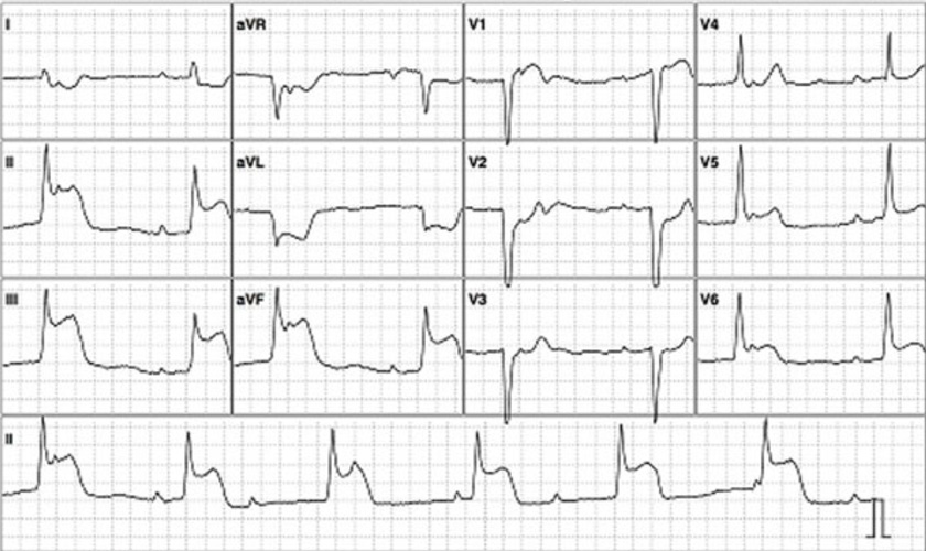
An image of a 12-lead ECG is provided below.

ECG systematic analysis
Although the paper recording speed and voltage calibration are not reported, we’ll assume it to be 25 mm/s, and 10 mm/mV, respectively (standard).
This ECG shows the following:
Rhythm: Complete AV block with a junctional escape rhythm
P waves: Normal axis and morphology, rate is 60 bpm
PR interval: Not defined (complete AV block)
QRS complex: Pathologic Q waves in lead V1-V3 and aVL. Normal duration, voltage and axis. Rate is 40 bpm.
QT interval: 440 ms (QTcB)
ST Segment:
ST elevation in leads II, III, aVF, V5-V6 and V1. Reciprocal ST depression in leads I and aVL.
There’s also ST depression in lead V2.
T waves: T wave inversion in leads I and aVL
Overall, this ECG demonstrates an inferior-lateral STEMI with complete AV block and a junctional escape rhythm.
Correct answer
The correct answer is ischaemia of the AV node due to RCA occlusion (RCA occlusion -> AV nodal ischaemia).
The patient presents with chest pain, hypotension, and bradycardia. The ECG shows ST elevation in leads II, III, and aVF, alongside reciprocal ST depression in I and aVL, which is characteristic of an inferior STEMI.
In the context of an inferior MI, the right coronary artery (RCA) is often involved, leading to ischaemia of the AV node. This is because the RCA supplies the AV node in around 70-80% of cases. This can result in a complete AV block, characterised by a dissociation between atrial and ventricular rhythms, as seen in the ECG.
Regarding the incorrect options:
Ischaemia of the AV node due to LCx occlusion is less likely because the left circumflex artery (LCx) supplies the AV node in only about 10% of individuals. While LCx occlusion can sometimes present as an inferior MI, it is less commonly associated with AV node ischaemia compared to RCA occlusion.
Ischaemia of the AV node due to LAD occlusion is unlikely in this scenario. The left anterior descending artery (LAD) typically supplies the anterior wall of the heart, and its occlusion is more commonly associated with anterior MI, not inferior MI. Anterior MI is more likely to cause infra-Hisian block rather than AV nodal block.
Ischaemia of the His-Purkinje system is more commonly associated with anterior MI, where the left anterior descending artery is involved, leading to infra-Hisian block. AV block associated with inferior MI is usually supra-Hisian in origin.
Ischaemia of the bundle branches is not consistent with the ECG findings. Bundle branch block is more commonly associated with anterior MI and would most likely show with a ventricular escape rhythm (not junctional as in this case).
Does this patient need a permanent pacemaker?
As per the 2023 ESC guidelines for the management of acute coronary syndromes, implantation of a permanent pacemaker (PPM) is recemmended when high-degree AV block does not resolve within at least 5 days after the myocardial infarction.
Additionally, these guidelines advise against pacing if high-degree AV block resolves after revascularisation or spontaneously. 1
Summary
Atrioventricular block, which can occur during myocardial infarction (MI), can range from a first-degree AV block that has minimal hemodynamic effects to a complete AV block that can lead to shock.
One of the reasons why an AV block can develop during an MI is increased vagal tone. However, it’s important to note that it can also be caused by hypoperfusion of significant structures in the conduction system, which is the primary focus of this challenge.
In a right dominant circulation (which is 70-80% of the population),2 the atrioventricular node is supplied by the right coronary artery.3 Knowing this correlation helps us understand the frequent connection between an inferior STEMI and an AV block.
2023 ESC Guidelines for the management of acute coronary syndromes
Robert A Byrne, Xavier Rossello, J J Coughlan, Emanuele Barbato, Colin Berry, Alaide Chieffo, Marc J Claeys, Gheorghe-Andrei Dan, Marc R Dweck, Mary Galbraith, Martine Gilard, Lynne Hinterbuchner, Ewa A Jankowska, Peter Jüni, Takeshi Kimura, Vijay Kunadian, Margret Leosdottir, Roberto Lorusso, Roberto F E Pedretti, Angelos G Rigopoulos, Maria Rubini Gimenez, Holger Thiele, Pascal Vranckx, Sven Wassmann, Nanette Kass Wenger, Borja Ibanez, ESC Scientific Document Group , 2023 ESC Guidelines for the management of acute coronary syndromes: Developed by the task force on the management of acute coronary syndromes of the European Society of Cardiology (ESC), European Heart Journal, Volume 44, Issue 38, 7 October 2023, Pages 3720–3826, https://doi.org/10.1093/eurheartj/ehad191
Shahoud JS, Ambalavanan M, Tivakaran VS. Cardiac Dominance. [Updated 2022 Sep 26]. In: StatPearls [Internet]. Treasure Island (FL): StatPearls Publishing; 2025 Jan-. Available from: https://www.ncbi.nlm.nih.gov/books/NBK537207/
Heaton J, Goyal A. Atrioventricular Node. [Updated 2023 Jul 25]. In: StatPearls [Internet]. Treasure Island (FL): StatPearls Publishing; 2025 Jan-. Available from: https://www.ncbi.nlm.nih.gov/books/NBK557664/

