
AV dissociation doesn't always mean AV block
There are three basic mechanisms by which the atria and ventricles beat independently, and only one of them is AV block.

AV association
Normally the atria depolarise first, and that impulse travels down through the AV junction (the AV node and His bundle) to trigger the ventricles. The ventricular beat happens because the atrial one did, and that causal link is atrioventricular (AV) association. It is what you confirm every time you check that a P wave sits in front of every QRS, and vice versa.
The name carries a direction, atria to ventricles. A link can also run the other way, ventricle to atria (ventriculoatrial), when a beat from below conducts back up and captures them. Either direction is association; dissociation is neither.
That link does not have to be one for one. In atrial flutter, for example, where the atrial rate is around 300 bpm, you could have every other atrial impulse travelling down to the ventricles, producing a ventricular rate of 300/2 = 150 bpm. That AV association would be called 2:1.
AV dissociation
When that link breaks, the two chambers carry on beating, but no longer because of each other. Each runs on its own pacemaker, on its own clock, and a given QRS no longer happens because of the P wave in front of it. Atria and ventricles beat independently. That independence is atrioventricular (AV) dissociation, and the way you get there is not always AV block.
Three basic mechanisms of AV dissociation
A subsidiary focus takes over the ventricles whenever it fires faster than the impulses arriving from above:
subsidiary focus rate > rate reaching the ventricles from above
There are three ways to make that happen: the first two leave conduction intact and simply outpace it, and only the third is a true conduction failure [1].
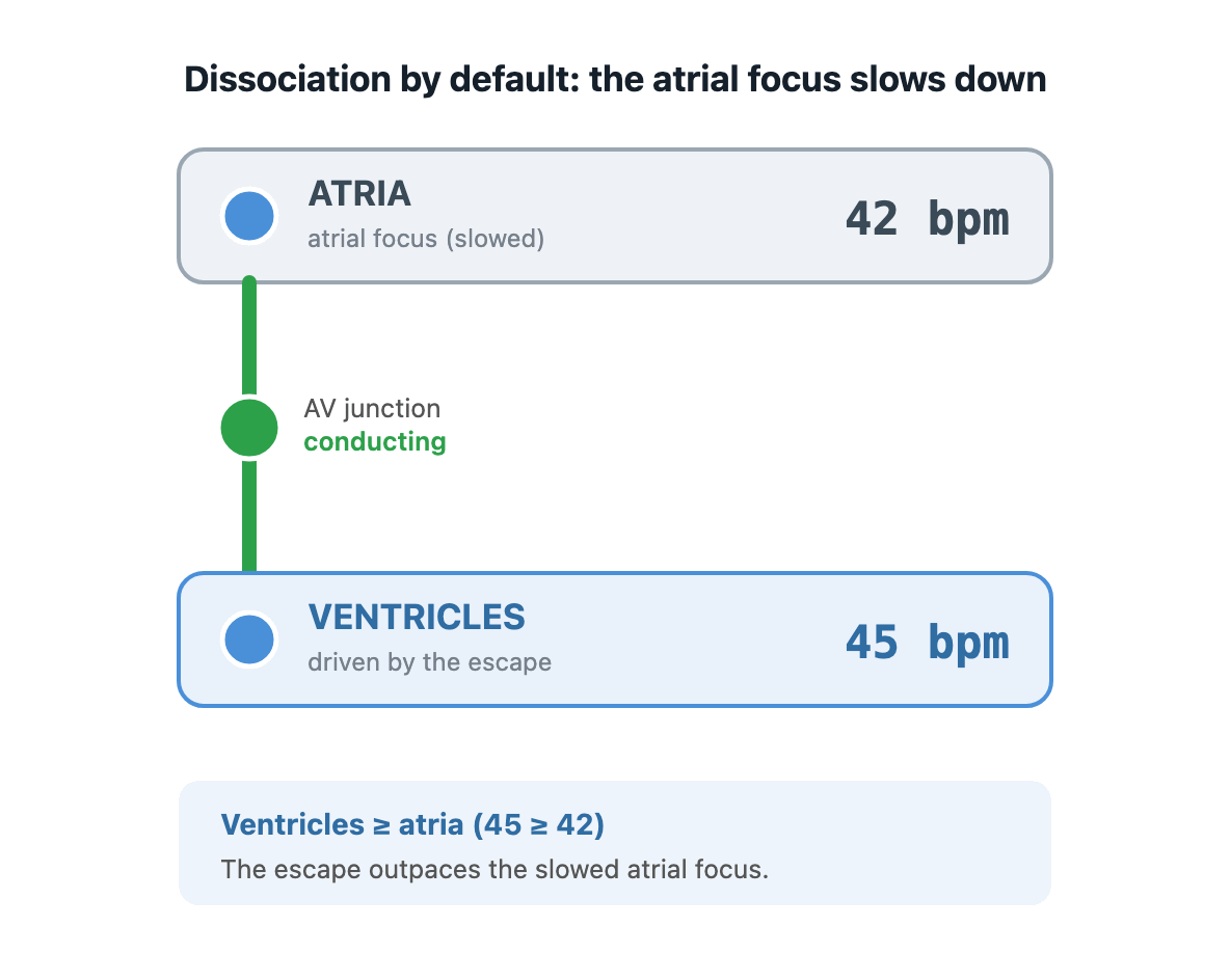
1. Decrease of atrial focus rate (dissociation by default)
Whatever the atrial focus is (normally the SA node), it has slowed down so much that it now beats more slowly than the intrinsic rate of a subsidiary focus below it.
That subsidiary focus now “escapes” to fill the gap: typically a junctional escape at 40-60 bpm, or a ventricular escape at 20-40 bpm.
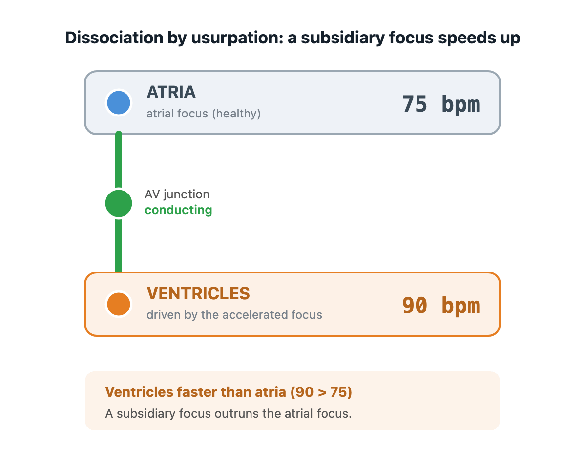
2. Increase of subsidiary focus rate (dissociation by usurpation)
A subsidiary focus accelerates past an atrial focus (which could have been a perfectly healthy SA node) and drives the ventricles.
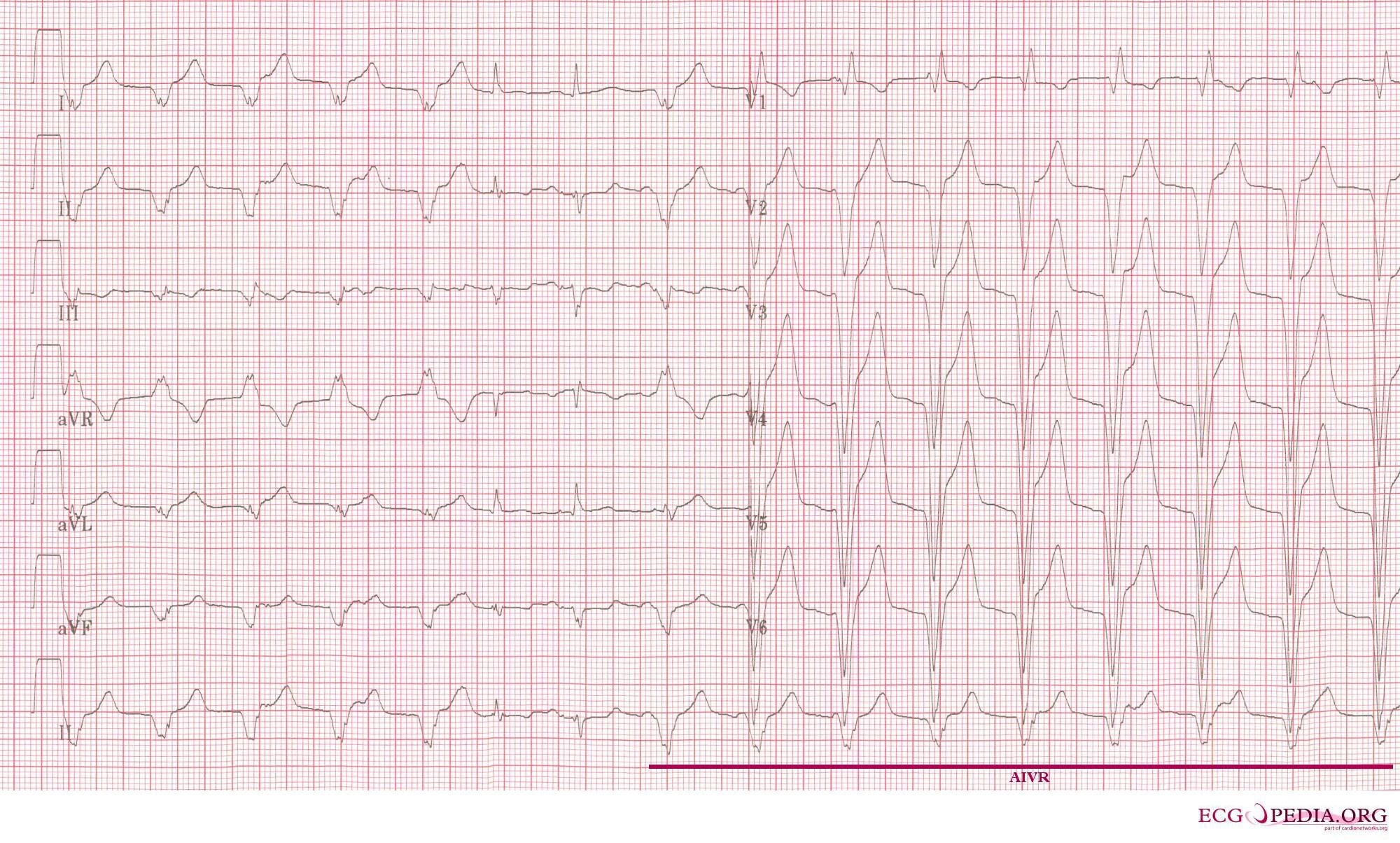
It might be an accelerated junctional rhythm, an accelerated idioventricular rhythm (60 to 100), or, at the dangerous end, ventricular tachycardia. Nothing needs to be blocked here: the AV junction may be perfectly healthy, just held refractory by the faster rhythm arriving from below [1].
This counts as dissociation only while the atria stay on a separate clock. If the fast focus also conducts back up and captures the atria (retrograde, ventriculoatrial conduction), the two chambers are linked again, upward this time, and that is association, not dissociation.
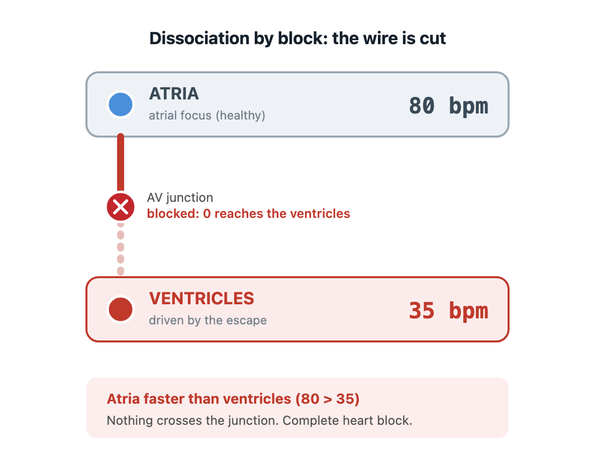
3. No atrioventricular conduction (dissociation by block)
In complete heart block nothing crosses the AV junction, so a subsidiary focus has to drive the ventricles or the patient arrests. The atria carry on at their own rate, the ventricles run slowly on an escape, and the two never meet.
When block is the cause, it is always third-degree (complete) AV block. First- and second-degree block still let some beats through, so they still tie the ventricles to the atria. Only when conduction fails completely do the chambers come fully apart. So a lot of AV dissociation involves no block at all, and the block that drives the chambers fully apart is only ever the complete sort.
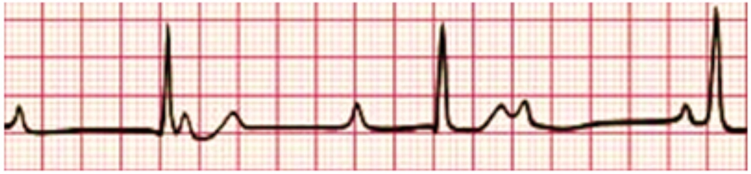
Block or no block: who is faster?
Look at the two rates and ask which one is faster.
Atrial rate > ventricular rate → block. Ventricular rate ≥ atrial rate → block not required.
The logic is mechanical. If the atria are faster and few or none of those fast atrial impulses are reaching the ventricles, something must be stopping them. The wire is cut, the ventricles are slow because they are surviving on an escape, and you are looking at complete (or possibly high-grade) heart block. This is the patient who may need pacing.
If the ventricles are the faster chamber, block is no longer the necessary explanation: they may be driven by an escape covering for a slow atrial focus, by an accelerated focus, or by VT, with the AV junction conducting perfectly [1]. But the rates only show that block isn’t needed to account for the rhythm, not that the junction is healthy. A fast subsidiary focus holds the junction refractory as each atrial impulse arrives, so it is never tested, and a complete block could be sitting underneath it whenever the subsidiary focus happens to outrun the atria.
So the label on its own does not get you far. “AV dissociation” tells you the atria and ventricles have come apart, but not why. And the mechanism is what decides what you do next.
Capture and fusion beats: is the wire still live?
In dissociation by default and by usurpation the AV junction still works; the subsidiary focus just happens to be in charge. So every now and then, when an atrial impulse arrives at a moment the AV junction is ready to accept it, it gets through.
A capture beat is that atrial impulse conducting all the way down the normal His-Purkinje system. It produces a QRS of the patient’s usual narrow shape, arriving early and briefly interrupting the dissociated rhythm.
A fusion beat is a collision: the atrial impulse and the subsidiary focus reach the ventricle at the same moment, each depolarising part of it, giving a hybrid QRS.
Their presence proves the wire is live. You cannot get a capture beat in complete heart block, because by definition nothing conducts. So a single capture or fusion beat tells you straight away that this is not third-degree block. And when the underlying rhythm is a broad-complex tachycardia, that same beat is close to a signature of ventricular tachycardia: proof that independent atrial activity exists and occasionally slips through [1]. It is one of the most useful clues for telling VT from an SVT conducted with aberrancy [2].
Summary
Independent P waves and QRS complexes are an observation, not a diagnosis. There are three ways to arrive at it:
By default: the atrial focus (usually the SA node) slows below a subsidiary focus, which escapes to fill the gap. The AV junction still conducts.
By usurpation: a subsidiary focus accelerates past a healthy atrial focus and drives the ventricles: an accelerated junctional rhythm, an accelerated idioventricular rhythm, or VT. The AV junction still conducts.
By block: conduction fails completely, so an escape has to drive the ventricles. This is the only one of the three that is genuine AV block, and it is always third-degree.
References
Rahman MFF, Yandrapalli S. Atrioventricular Dissociation. [Updated 2023 Jul 24]. In: StatPearls [Internet]. Treasure Island (FL): StatPearls Publishing; 2026 Jan-. Available from: https://www.ncbi.nlm.nih.gov/books/NBK563205/
Wellens HJ. Electrophysiology: Ventricular tachycardia: diagnosis of broad QRS complex tachycardia. Heart. 2001 Nov;86(5):579-85. doi: 10.1136/heart.86.5.579. PMID: 11602560; PMCID: PMC1729977.