


What is the rhythm?
Rhythm recognition always requires a careful and systematic analysis of an ECG.


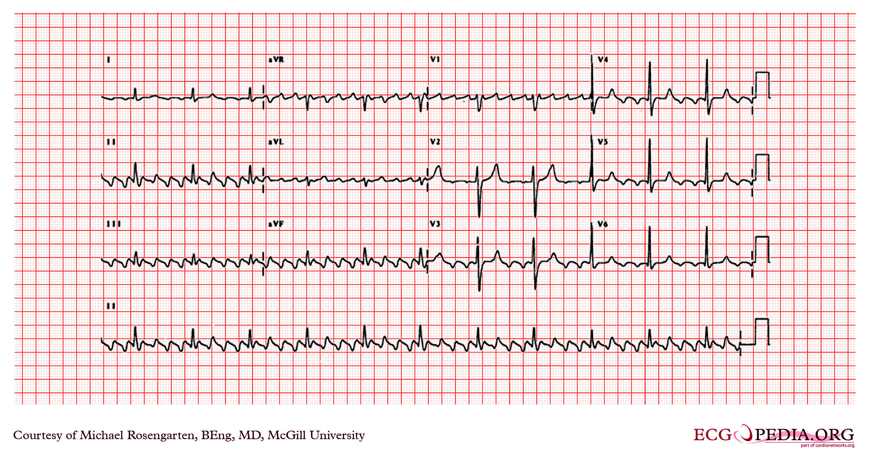
Figure explanation
The ECG in the figure above (1) was interpreted by a computer algorithm as atrial flutter. However, on careful examination, this is not the correct diagnosis.
The actual rhythm is sinus rhythm (2), and we know that by looking at lead I.
This ECG shows sinus rhythm with artefacts caused by patient movement. The computer misread these artefacts as flutter waves, but the true diagnosis can be uncovered with a careful, systematic approach.
Lead I is key in solving this puzzle. Unlike the inferior leads, lead I shows a normal sinus rhythm without the presence of artefacts.
Lead I records the potential difference between the left arm and right arm, meaning the left leg is not involved in this lead's measurement. Therefore, the absence of artefacts in lead I indicates that the source of the artefacts is likely related to movement of the left leg.
How to avoid misinterpretation
Take your time.
I believe there is one main reason why someone would misinterpret this ECG: by not taking enough time to interpret it.
We need to be patient when interpreting ECGs; we need to take our time. ECGs provide a wealth of data, not only about the heart but also about other aspects, such as evidence of intracranial hypertension, electrolyte abnormalities, ethnicity, physical activity levels, and age.
Always compare leads. However, to get to that point, we need to spend some time looking at the ECG.
An ECG with atrial flutter for comparison
Let’s compare leads:

We can see the following:
Consistent regular atrial activity with a rate of around 300 bpm (5 mm) present in all leads
A sawtooth pattern of atrial waves (i.e. flutter waves)
1 QRS conducted every 4 flutter waves
Therefore, this is atrial flutter with 4:1 ventricular conduction.
Notes
(First image) Erroneous computer-based interpretations of atrial fibrillation and atrial flutter in a Swedish primary health care setting - Scientific Figure on ResearchGate. Available from: https://www.researchgate.net/figure/ECG-interpreted-as-atrial-flutter-Artifacts-due-to-patient-movement-are-present_fig1_337044786 [accessed 24 Oct 2024]
Why most likely sinus rhythm: because we need to know that the P wave axis is normal as well, and we can only really see P waves in lead I. So, if the P wave was upright in lead aVF as well, then it would be even more likely that this is sinus rhythm.