When every second counts | ECG challenge #1
Spot this ECG pattern fast—or risk missing a fatal diagnosis.
Clinical case
A 65-year-old man was admitted to the Coronary Care Unit with severe chest pain and dyspnoea. He has a history of previous myocardial infarction and coronary artery bypass graft surgery. He also has a history of hypertension and high cholesterol levels.
His blood pressure is 85/60 mmHg, his pulse is 110 beats per minute, and his respiratory rate is 30 breaths per minute. Oxygen saturation is 82% on room air.
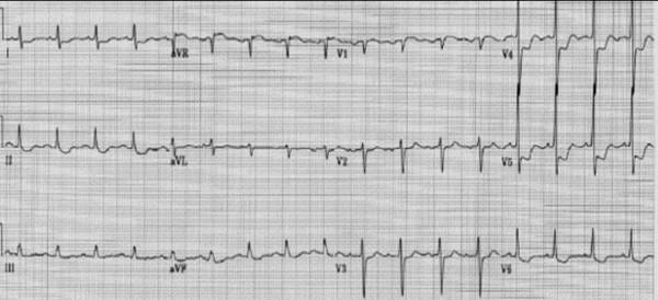
An image of a 12-lead ECG is provided below.
ECG systematic analysis
Although the paper recording speed and voltage calibration are not reported, we’ll assume it to be 25 mm/s, and 10 mm/mV, respectively (standard).
This ECG shows the following:
Rhythm: Sinus tachycardia at 107 bpm
P waves: Normal axis and morphology
PR interval: Normal (180 ms)
QRS complex: Normal morphology, axis and duration
QT interval: Slightly prolonged (~480 ms QTc)
ST Segment:
ST elevation in leads aVR and V1 (greater in aVR than in V1)
Widespread ST depression in leads V3-V6, I, II, and aVF
T waves: Biphasic in V4-V6
Correct answer
The correct answer is left main stem (LMS) obstruction.
The ECG reveals sinus tachycardia with ST elevation in lead aVR and V1, along with widespread ST depression in leads V3-V6, I, II, and aVF. This pattern is indicative of multivessel ischaemia or left main coronary artery obstruction.
.
ST depression ≥1 mm in ≥6 surface leads
AND
ST elevation in aVR and/or V1
→ THINK: Multivessel ischaemia or LMS obstruction
(Especially if the patient is hypotensive or tachycardic)1
(2023 ESC guidelines for the management of acute coronary syndromes)
.
Other options, such as acute anterior or inferior myocardial infarction, typically present with more localised ST elevation patterns. Pericarditis often shows widespread ST elevation without reciprocal depression, and pulmonary embolism may present with sinus tachycardia and right heart strain but not this specific ECG pattern.
If you suspect LMS or multivessel occlusion, your patient needs immediate cardiology attention!
Summary
In acute coronary syndromes, when you spot:
≥1 mm ST depression in ≥6 leads
ST elevation particularly in aVR (often greater than V1)
Think immediately: Left main stem (LMS) obstruction or severe multivessel ischemia.
Robert A Byrne, Xavier Rossello, J J Coughlan, Emanuele Barbato, Colin Berry, Alaide Chieffo, Marc J Claeys, Gheorghe-Andrei Dan, Marc R Dweck, Mary Galbraith, Martine Gilard, Lynne Hinterbuchner, Ewa A Jankowska, Peter Jüni, Takeshi Kimura, Vijay Kunadian, Margret Leosdottir, Roberto Lorusso, Roberto F E Pedretti, Angelos G Rigopoulos, Maria Rubini Gimenez, Holger Thiele, Pascal Vranckx, Sven Wassmann, Nanette Kass Wenger, Borja Ibanez, ESC Scientific Document Group , 2023 ESC Guidelines for the management of acute coronary syndromes: Developed by the task force on the management of acute coronary syndromes of the European Society of Cardiology (ESC), European Heart Journal, Volume 44, Issue 38, 7 October 2023, Pages 3720–3826, https://doi.org/10.1093/eurheartj/ehad191


Excellent way to "make it stick". Thank you!